
[email protected]
Medical Device
Research Infrastructure (ORIP)
UT - 1
3Helix
What do arthritis, osteoporosis, and some cancers have in common? They all show signs of tissue damage early on in their disease progression. But researchers and clinicians don’t have an easy way to detect this damage. If they did, diseases could be diagnosed earlier and more accurately.
The company 3Helix is trying to change that. When Johns Hopkins researchers Yang Li and Michael Yu were studying collagen—the main protein that gives tissues structure—the two founders accidentally discovered a way to identify damaged collagen. They started 3Helix in 2015 after moving to the University of Utah to develop their technology into a tool for studying diseases associated with damaged collagen.
3Helix is developing a method to visualize damaged collagen resulting from fibrosis and then associating the amount of damage to a level of disease severity with the goal of giving pathologists a more objective method of scoring disease progression.
Today 3Helix provides a fluorescent probe for researchers to highlight the damaged collagen in tissue samples, which can then be viewed and studied under a microscope. In 2016, the company received a Phase I Small Business Innovative Research (SBIR) grant from the NIH, which Chief Technology Officer Lucas Bennink says allowed them to flesh out their research, convert it into a useful product, and start selling it in 2017. In a two-year span, the company increased their sales by almost 3,000 percent.
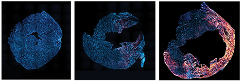 3Helix's fluorescent probe can detect the progression of damage caused to the tissue surrounding the heart after three days (middle) and seven days (right) after a heart attack compared to a normal sample (left) by revealing the amount of damaged collagen.
Bennink says their 2019 Phase II SBIR has given 3Helix the ability to direct their efforts toward specific diseases. The company is now undergoing studies on liver and kidney fibrosis—scarring that results from injury or long-lasting conditions—to develop a new way of diagnosing disease severity. Currently pathologists rate fibrosis severity through a grading system that can be subjective. 3Helix is developing a method to visualize damaged collagen resulting from fibrosis and then associating the amount of damage to a level of disease severity with the goal of giving pathologists a more objective method of scoring disease progression. The company is currently trying to apply this method to the degenerative joint disease osteoarthritis.
Today 3Helix has sold their product to more than 100 users in at least 10 countries since making their product available. Researchers have published more than 50 research papers from results acquired using their product.
Bennink says they have gained both recognition and validation from receiving the SBIR grants. “We wouldn't be where we are without [NIH] support.”
Along with using their product as a diagnostic tool, 3Helix also plans to explore its use as a therapy and to monitor disease progression. “It has the potential to have a huge influence on multiple different disease states,” says Bennink.